The basic ultrasound machine uses a series of transducers to create images of your body. The images produced by this machine can be divided into two main modes: the B-mode, which is also known as a 2D mode, and the M-mode, which is known as a motion mode. In both modes, a linear array of transducers scans a plane through the body. During the B-mode scan, an image is produced, while M-mode images and videos are produced through the successive pulses of the transducers. These images show organ boundaries moving relative to the probe, which can help determine the velocity of specific organ structures.
Doppler
Doppler ultrasound is a type of diagnostic test that uses reflected sound waves to create images of organs, soft tissue, and blood flow. This type of test is safe and does not expose patients to any radiation, making it the preferred method of imaging for pregnant women and unborn children. There are several types of Doppler ultrasound exams available at ultrasound centers. One type can help physicians determine if an angioplasty is appropriate. Doppler ultrasounds can be done on the entire body or only selected parts. Before undergoing a test, patients must be fasted for at least 12 hours and must refrain from using tobacco, alcohol, or other nicotine products. Nicotine narrows blood vessels and may affect the results of the test. The procedure usually takes 30 to 60 minutes and requires the patient to lie still.
Using high-frequency sound waves, Doppler ultrasounds can measure the blood flow through the veins and arteries of the arm, leg, or other part of the body. This is an excellent way to detect circulatory problems and diagnose them early. However, doppler ultrasounds should only be performed in a vascular medicine department of a hospital, where a physician is trained in this procedure.
Bi-planar
Several studies have been conducted to determine the best way to use ultrasound in medical diagnostics. Using a bi-planar ultrasound, for example, can be beneficial for the diagnosis of some spinal conditions. The most common method for assessing the health of the spine is through a CT scan. The scan can be performed in either a seated or standing position.
Ultrasound scans can also be used for research. Researchers have used ultrasound to study blood flow and muscle movement. They can also use the images to look at heartbeats and fetal movement.
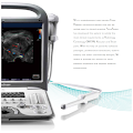
Transducer probe
An ultrasound machine uses high-frequency sound waves to examine tissue inside the human body. The transducer is positioned perpendicular to the target nerve or tissue. Because soft tissues do not have much difference in acoustic impedance, very little sound is reflected by them.
The ultrasound machine consists of a computer console, a video monitor, and a small microphone called a transducer. The transducer sends high-frequency sound waves and listens for their echoes. The principle is similar to that of submarine sonar.
Gel application
Ultrasound gel can be used in non-invasive ultrasound procedures and therapeutic ultrasound procedures. Its high viscosity provides good acoustic coupling with tissue, and its viscous nature ensures that it remains in place during a procedure. Ultrasound gels are also useful for their lubricant properties, which make them useful for imaging and measuring procedures. Ultrasound gels are also biocompatible, food-grade, and sterile.
Gels can also be used in dental applications. They can be used to improve the comfort of dentures, and they may also be used to treat oral organs. Gels are also used to enhance the imaging and therapeutic capabilities of intra-oral ultrasonography and dental ultrasound. Gels are compatible with ultrasound transducer equipment, and they can easily be integrated into new ultrasonic dental imaging systems.
Biological effects of non-thermal origin on ultrasound waves
The biological effects of ultrasound waves on humans are largely unknown. Although a few studies have documented the presence of some adverse effects, others have not. The intensity and frequency of ultrasound waves are measured in joules (J) and the power is measured in watts (W). Intensity is a function of area unit (cm2), and the power is measured as mW/cm2. These are the conventional measures of ultrasound intensity.
One study examined the impact of ultrasound on lung tissue in a rabbit. It used pulsed-wave ultrasound to damage lung tissue. Another study involved heating of bone tissue by ultrasonic waves.
| Similar Content |
|---|
| What is an Ultrasound Machine Used For? |
| 5 Uses of Ultrasound |
| What Can an Ultrasound Test Detect? |
| How Is Your Ultrasound Machine Working? Part 2 |